Showing posts with label The Doctor's Bag. Show all posts

Thursday, April 26, 2018

We'd Better Open his Head!


THE DOCTOR'S BAG

The blog about 19th Century Medicine and Surgery

By Keith Souter aka Clay More


TREPHINATION AND HEAD INJURIES


Last week, knowing my interest in the history of medicine, Jacquie Rogers drew my attention to a post on Facebook about a 5,000 year old cow's skull from an excavation in France. It had a surgically created hole in the right side. No bone healing had taken place, so it was suggested that this was either an early attempt at veterinary surgery, or it was a practice attempt by an ancient surgeon before attempting a trephination on a human, possibly after a head injury.

Head injuries are common in tales about the Old West. That is not surprising, considering all those gunfights, fist-fights and falls from horses. The town doctor on the frontier would probably have to be adept at treating them. That could include trephining the skull - effectively, opening the head.


Treatment of a skull injury from Fieldbook of Medicine 1517

Nowadays we have CT and MRI scans which can give us sophisticated images of the body. A CT scan stands for Computerised Tomography, which involves taking x-rays of the brain from various angles, which are analysed by a computer to build a 3-d image of the brain. This will show any fracture or haemorrhage. A MRI scan stands for Magnetic Resonance Imaging. It uses magnetism, ultrasound and computerised technology to build up images of the inside of the body.  These can show the tissues and any abnormalities in surprising detail.

Back in the Old West there were no such luxuries. X-rays were only discovered by William Roentgen in 1895. The first use of them diagnostically only started the year after when Dr John Hall-Edwards in Birmingham, England started to use the technique. The town doctor had to use his clinical skills.

The unconscious patient
The doctor would assess the patient, by examining the scalp for a wound or local bruising. He would examine the ears and nostrils for bleeding, both of which could indicate a significant head injury, with fracture of skull bones.

Coma is the state of absolute unconsciousness when the patient does not respond to any stimulus. Squeezing the ear lobe or using the knuckles of the hand to rub over the sternum (breast bone) are extremely painful and usually will evoke a response in someone who is in a semi-coma. This means that they only respond to painful stimuli. A full coma patient will not even respond to pain.

The pulse would be taken frequently, say every 15 minutes in an unconscious patient. A slowing of the pulse is called bradycardia and may indicate internal hemorrhage somewhere, possibly inside the skull.

The semi-conscious patient
The patient may well be confused, so the level of confuse would be assessed. A rule of thumb gives mild, moderate or severe states of confusion.

Mild - some coherent conversation  is possible
Moderate - out of touch generally, but will answer with name or occupation
Severe - no sensible answers given, but will respond to simple commands such as hold my hand.

Deepening confusion may indicate hemorrhage. Sudden vomiting may also be highly significant.

Examination of the cranial nerves
There are twelve paired nerves which come directly out of the brain to supply the head and neck and some of the internal organs. These are separate from most other nerves, which come out from the spinal cord.

1st nerve - olfactory nerve - sense of smell
2nd nerve - optic nerve - vision
3rd, fourth and sixth nerves - occuolomotor, trochlear and abducent nerves move the eye and operate the pupils - get the patient to follow the finger and also shine a light in the eyes - the pupils should constrict
5th nerve - trigeminal nerve - sensory to the face and operates the masseter muscles, the large muscle that moves the jaw - tested by asking to clench the jaw
7th, facial nerve - movements of the face - ester by asking to see the teeth
8th, acoustic nerve - for hearing. Can he hear?
9th glossopharyngeal nerve - to the pharynx - cannot be tested
10th vagus nerve - multiple internal functions, but also moves the palate - tested by asking the patient to say 'Ah.' The palate should move when he does
11th nerve - accessory nerve - tested by asking to shrug the shoulders
12th nerve - hypoglossal nerve - moves the tongue.

Significantly the movements should be equal on both sides. One sided results could indicate a problem with one side of the brain.

Hutchinson's pupils
These are a set of guidelines devised by Sir Jonathan Hutchinson (1823-1913), a professor of Surgery at the Royal College of Surgeons of London. He wrote a ten volume  Archives of Surgery between 1885 and 1899.


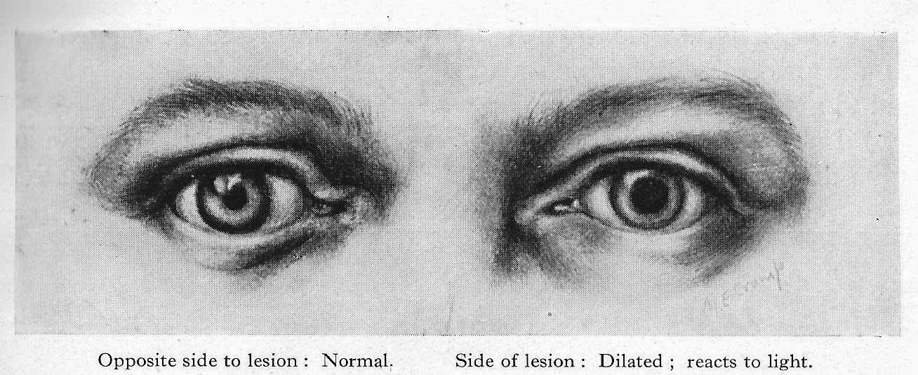
Hutchinson's pupil, described by him indicates that an intracranial mass (a tumour or blood clot) will result in a fixed (meaning it will not change size even when light is shone on the eye) and  dilated pupil on the same side. This is very significant, since it would indicate where to make the trephination.


Racoon eyes and Battle's sign
Basal skull fractures, that is, fractures of the bones that make up the base of the skull, including the temporal bone, occipital bone, ethmoid and sphenoid bones, can be associated with particular bruising.

Racoon eyes

Racoon eyes, also known in the UK as Panda eyes, can occur several hours after a basal skull fracture. they look like black eyes from direct trauma, but the bruising is limited to the orbit. There are other causes, but in the context of head injuries in your fiction writing, be aware of this sign.

 Battle's sign, named after the English surgeon William Henry Battle (1855-1936) is bruising that occur can  behind the ear, again some hours after a basal fracture. Like the racoon eyes it is not necessarily a sign of trauma where it appears, but a sign of a basal skull fracture.

Battle's sign  

Your frontier doctor might not have known of them by those names, but he may well have known from past experience, or war service, that they could be of significance.


General physical examination
Here the focus is on assessing the power of each limb and comparing one see of the body with the other. Disparity between the sides could indicate developing or developed paralysis.

Also testing the reflexes with a 'patella or tendon hammer.' The following reflexes are tested:

Biceps tendon by tapping the tendon in the hollow of the elbow.
Triceps tendon by tapping the tendon on the back of the elbow when the elbow is flexed.
Patella tendon by tapping underneath the stella (kneecap) with the leg flexed.
Achilles tendon  by tapping the tendon at the back of the ankle.

Lack of reflexes on one side would be significant.

Trephination
The words trephination and trepanation are used interchangeably, since both come from the Greek trypanon, meaning 'to bore.' Archaeological evidence shows that trephination, the boring of a hole in the skull was used in early tribal societies. It was presumably thought that this would let out evil spirits. Examination of many skulls which have been trepanned in this way shows that healing of bone around the site of the boring took place, indicating that in many cases the operation was a success. Incredibly, they used three methods - cutting, scraping and drilling.

The oldest trepanned skull was found at a neolithic burial site at Ensisheim in France. It has been dated to 7,000 years ago.

The reason that it could have helped is by releasing the pressure upon the brain, which would follow a hemorrhage. Unfortunately, in those people who were not suffering from a rise in pressure it may have done actual harm.

The ancient Egyptians had actually developed a quite sophisticated system of medicine and surgery with doctors who specialised in one area of the body. thus they had eye doctors, stomach doctors and head surgeons.

The Edwin Smith papyrus, written in about 1500 BC, is essentially an ancient Egyptian textbook of surgery. It describes surgical instruments and techniques and discusses 48 cases of injuries, including head injuries.


The Edwin Smith Papyrus c 1500 BC - an ancient Egyptian textbook of surgery

A beautiful description of ancient Egyptian surgery is given in the 1945 historical novel The Egyptian by the Finnish writer Mika Waltari, which became an international bestseller and later a Hollywood blockbuster in 1954. In the novel the main character Sinuhe, who became royal physician to Pharaoh Akhenaten (father of Tutankhamen), is apprenticed to Ptahor, the 'opener of head.' He shows him how to examine a patient and diagnose where there may have been a problem in the head from an assessment of the state of consciousness and the use of the limbs. He then shows him how to remove a piece of skull and replace it with a silver plate which is bound with bandages while they await recovery. 


Ancient Greek surgical instruments. Note the trephine in diagram 'a' with a central pin

The technique of trephination was also used by the Greeks and the Romans. The instruments required became increasingly sophisticated.

We're going to have to open his head
The experience of British surgeons with  gunshot winds to the head during the the Crimean War (1853-1856) was not promising. Trephination was associated with an extremely high mortality of over 95 per cent. George H B Macleod, the chief surgeon of the British Expeditionary Force advise against the operation.

During the American Civil War the operation was performed with better results. The survival rate improved to more than 20 per cent. Then after Lister's aseptic techniques were accepted, recovery rates continued to improve.

In 1882 Samuel W Gross wrote a textbook A System of Surgery, in which he quoted a 41 per cent recovery rate after gunshot trephination. 

The trephination operation
The instruments needed:

Trephine - a cutting instrument with a cylindrical blade - usually with a cutting circle of one inch


An antique trephine with horn handle

Chisel and mallet


Rongeur - a strong forceps for grasping bone


Also needed would be a scalpel and various forceps for holding tissues, a gouge for smoothing roughed or fractured bone edges and an elevator to lift the bone disc that was cut.

The operation
The patient's head would be shaved and washed. After Lister this would have been with carbolic soap and then it would be scrubbed and washed with a 1 in 20 solution of carbolic acid in alcohol. 

The head would be supported on a sandbag and sterilised towels applied around the site to open the skull. In the earlier frontier days, this would not have been thought necessary.

When a wound already exists and a fractured spicule was apparent, the cite would be exposed by enlarging the wound. When the scalp was not wounded, as in a clubbing head injury or from a fall, then a semilunar flap of skin would be cut and raised. It would be cut so that the free end would point downwards. It would be a shallow curve, carefully made to avoid the main scalp arteries.

Alternatively, a V-shaped incision could be made, again with the V pointing downwards. It should be so arranged as to allow free-draining of blood. 

The incision should be carried down to the bone and the tissues helped with forceps. Then the flap would be turned upwards. A suture would be placed through it so that the flap could be held out of the operative territory.

Any bleeding vessels would be secured with pressure forceps to close the vessels off. 

Any spicules of fractured bone could be chiseled off with the chisel and mallet. 

If there is no fracture, then the circular trephine is applied. The central pin could be bored into the bone, then the trephine is made to cut into the skull by light, sharp movements from left to right and from right to left.

At first bone dust is dry, but it soon becomes soft and bloody. Once through the bone, the pressure changes dramatically. 

The area must be kept free of bone dust by irrigation with saline. 

The trephine is then gently rocked back and forth to allow it to move, then the elevator is inserted. The  ronguer forceps can then be applied carefully to remove the disc. 

The blood  or the blood clot can then be removed. It may well express itself. The area needs to be irrigated and any bone spicules or bone dust removed. 

The bone disc is placed in a china cup and soaked with warm (sterilised) water, ready to be replaced.

Once replaced the skin flap is brought back into place with silkworm-gut sutures.

In later years a spiral rubber tube could be used to drain between the sutures. 

It wasn't always a head injury
Abscesses could also cause raised intracranial pressure and need trephination.  


Choosing the site of trephination was important, since you need to avoid the middle meningeal artery.  The diagram shows the areas that were commonly used. They would avoid the area between A and B, since the artery runs underneath the cranium. Points A and B would be used if a middle meningeal artery haemorrhage was suspected.

This sort of hemorrhage can cause an epidural haematoma. This is a blood clot forming between the dura mater (the thick outer layer of the meninges, the membranes that cover the brain) and the cranium. Rupture of the middle meningeal artery is often the cause.  It comes on after a head injury when someone is knocked unconscious. They then recover, seem to regain lucidity and even go off and resume normal activities. Later on they very quickly go drowsy and lose into unconsciousness s the clot forms. It is a life-threatening event and the trephination could be life-saving.


Finally, a case!
Report of a Case of Fracture of the Skull treated by trephining - by Ira Perry, Assistant Surgeon, US Army 

Jesus Soldaeo, a Mexican, while at a drunken revel in Brownsville, Texas, June 1, 1866, was struck wit the stock of a gun, which used extensive contusion and fracture of the skull from the external angle of the orbit of the left eye toward the occiput. the fracture could be traced through the scalp  distance of six inches........The patient as admitted to the post hospital totally unconscious, with slow pulse and very feeble. The case being considered hopeless, water dressings only were applied....on the third day, the patient being still alive, the trephine was applied, and fragments of bone elevated; three days later the patient was slightly conscious....Until the tenth day the patient had taken nothing but cold water, and now was given some gruel; bowels were opened for the first time.....On June 25th consciousness returned completely and a gradual improvement took place.  July 17th the wound was almost healed, and the patient was removed fro the hospital by his friends. 


Be bold!
I have used trephination in some of my western stories. It is tough surgery performed in life and death situations, so the operator has to be bold. Which of course, all of us as western writers have to be.

***


Thursday, March 22, 2018

THE DOCTOR'S BAG


The blog about medical and surgical practice in the Old West

by Keith Souter 

aka CLAY MORE


MORE NOTES ON ARROW WOUNDS

Welcome back to the days of digging out arrows, bullets, mending busted bones and removing tonsils on a well-scrubbed kitchen table. After I complied two years or so of blogs into the book The Doctor's Bag, published by the good folk at Sundown Press, I thought that I had probably said as much as I needed to on the subject. Yet on looking back at some of those articles and the sources I used, I think there is quite a lot more information that writers of westerns and historical novels might find useful.

This month I am revisiting one of the earliest articles that I wrote for The Doctor's Bag. It was entitled Dig It Out, Doc! Part one - Arrow wounds.  It covers the history of arrow wounds from antiquity until the work of Doctor Joseph H. Bill. Of necessity, it merely covered the basics about the treatment of arrow wounds. There is certainly all that the writer needs in the chapter in the book if  he or she needs to deal with such a scene in their novel or story.  In this article I thought I'd look at one or two cases that Joseph Bill used to illustrate his work and also give some of his views about prognosis. 


A contemporary drawing of Bradmore's arrow extractor, from the book, Fair Book of Surgery, c 1450

Doctor Bill was an Assistant Surgeon in the US Army and had great practical experience in the treatment of arrow wounds. He wrote a short treatise entitled Notes on Arrow Wounds published in the American Journal of Medical Sciences, October, 1862.

The writing of such texts has changed considerably over the years. Doctor Bill would give his opinions  about the dangers posed by the different tribes, when discussing the different types of arrows. He writes:

"Before long these wounds will become of unfrequent occurrence, for our Indian tribes are fast being exterminated. We propose, in the first place, as a matter of historical interest, to state in this article that we know of arrow wounds. The subject still presents much of practical interest to the surgeon, and must continue so to do, in a greater or less degree, for the future."

Don't just pull it out!
He gives an account of the way that arrows are made, which he indicates is crucial to understanding the aims of treatment.  He gives this scenario:

"Let us suppose a case to illustrate and explain our meaning. An arrow is shot at a man at a distance of fifty yards. It penetrates his abdomen, and without wounding an intestine or a great vessel, lodges in the body of one of the vertebrae. The arrow is grasped by the shaft by some officious friends, and after a little tagging is pulled out. We said the arrow is pulled out. That was a mistake, it is the shaft only of an arrow that is pulled out. The angular and jagged head has been left buried in the bone to kill - for so it surely will - the victim. The explanation of such mishaps isthmus: the ribbon of tendon which compressed together the slit ends of the end of the arrow, and so clamped the head and the shaft together, had become wetted with the fluids effused in the course of the wound. When wetted, it was, of course, lengthened, and, if lengthened, loosened. It ceased longer to bind together the split sides of the shaft; this and the head were, consequently, very feebly united and readily detached. Experience has abundantly shown, and none know the fact better than the Indians themselves, that any arrow wound of chest or abdomen in which the arrow -head is detached from the shaft and lodged, is mortal. 

From this we conclude that the danger peculiar to all arrow wounds is, that the shaft becoming detached from the head of an implanted arrow, leaves this so deeply embedded a bone that it cannot be withdrawn, and that, remaining, it kills."

So there you have the stark fact. All those novels and movies where the arrow was plucked out and cast aside, had actually consigned the person to death, rather than a short convalescence after whiskey was poured over the wound and it was covered with a pad and bandage.

Laudable pus 
Pus is the white or yellow inflammatory fluid that forms when infection is present. Its colour depends upon its constituents, or which bacteria are present. Surgeons from the days of antiquity talked about 'laudable pus.' They thought that pus was a natural part of the healing process and that it was a necessary nuisance. It was not until the Germ theory  that they realized it was as sign of infection.

One of the oldest surgical maxims is 'Ubi pus, ubi evacuation,' which is Latin for 'where there is pus, let it out.'

Doctor Bill was working in the days before the Germ Theory, but he was aware of the special problems that arrow wounds caused, even after the whole arrow was removed.  He noted that the muscular tissue would contract after removal, distorting the tract of the arrow. Thus, when pus formed, it would lose its natural channel and collect in a pocket within the wound. Effectively it would become an abscess. The next stage was often sepsis, or spread of the infection to the bloodstream, often producing death in days or weeks. 

Poison arrows
Despite the problem of infection, Bill said that some tribes deliberately poisoned their arrows. 

"It is occasionally the practice of some of our Indian tribes to poison their arrows. The plan pursued is this (on the testimony of a Moquis Indian). The liver of some animal is exposed, and a rattlesnake compelled to insert his fang into it. The animal is at once killed, the liver removed, and wrapped up in the skin and buried. After seven or eight days the bundle is dug up, and the arrow-heads dipped in the pulpy and putrescent mass inclosed in the skin. After they are dry they are dipped in blood, again dried, and preserved for us."


Sometimes people did survive chest wounds
This is recorded as CASE V:

A Mexican, name unknown, whilst travelling the road near Fort Defiance, was shot by Navajoes with five arrows, all the arrows injuring the lung, and one of them passing through the upper border of the liver. I saw the man twenty minutes after the accident. The bleeding was most profuse, and the man fainted. Both lungs were wounded, I made the worst possible prognosis, but proceeded to extract the arrows, all of which I  safely removed. After the haemorrhage had ceased, I applied muslin soaked in collodion to each of the wounds, eight in number (some had penetrated right through the chest) and had the patient put to bed, and given  gram of morphia.

The next morning the patient was more comfortable than I had expected. He had been vomiting, and I gave him some acetum spit and acetate of lead, to quiet this and restrain secretion. In the afternoon the man complained of pain the right side, and difficulty in breathing. I removed the dressing from the wound, involving the liver and lung, and broke up some clots whereupon a considerably quantity of mixed fluids were discharged from the wound. These contained bile as I discovered by Pettenkofer's test (a standard test for bile)  . This procedure greatly relieved him, and he expressed himself much better. I ordered the prescriptions of the morning to be repeated.

The next day the patient vomited a considerable quantity of black, decomposed blood, and complained if the frequency of his urination. Urine very dark coloured, sp gravity 1036; urine contained chalice acid and sugar. All medicines ordered to be discontinued. 

After this the patient gradually recovered, all the wounds healing by first intention, except that implicating the liver, and that ceased discharging on the sixth day. In two weeks the man left the hospital, his urine still containing sugar, but no bile. In a few days the lat traces of sugar disappeared from his urine. He recovered completely.

Protection advocated
Doctor Bill felt that more should be done to protect soldiers from arrow wounds. 

"We wish in conclusion to recommend to those in authority the plan for protecting soldiers and others exposed arrow wounds with a slight curios. The Indians have a method of dressing bulls' hide for shields for themselves which renders it arrow proof. A cuirass made of such material, protecting the whole trunk from  before and behind, need not weigh more than eight or ten pounds, and by means of it a soldier could enter an Indian fight with fair chance of escaping death."


___________________


Thursday, April 27, 2017

THE HEALING STONES OF ST FILLAN


THE DOCTOR'S BAG

the blog about the medicine and surgery of yesteryear

Keith Souter aka Clay More


This month I am not going to talk about the medicine of the Old West. Instead, I am going to mention the 'old medicine' of the West of Scotland.

Recently I paid a visit to an elderly relative in Carnoustie, which is in the east coast,  before heading across country to spend a couple of days hiking in Glencoe. On the way across I made a detour to stop off at the beautiful little village of Killin. It is situated astride the River Dochart with the Falls of Dochart running through the village. 


The Falls of Dochart


The Healing stones of St Fillan
My purpose was to visit the Breadalbane Folklore Centre which is housed in the old mill on the bank of the river. The original mill was apparently built by St Fillan. Inside the mill are the famous Healing Stones of St Fillan.

           
St Fillan was a monk from a noble Irish family, a follower of St Columba, who came to Killin in the seventh century to try to convert the Picts from their ancient pagan ways. His name means wolf cub, or little wolf.There are many legends that have grown up around him and they have drifted into the folklore of Scotland. He was said to have been born with a stone in his mouth. His father threw him into a lake, but angels looked after him until he was found and cared for by Bishop Ibar. He brought him up as his son in the  Christian faith. 

So much for legend. It is known that he taught in the vicinity, operated a meal mill and worked as a healer.
           
In his healing work he used stones that had been gathered from the river bed. There were eight of them and he bequeathed them to the community, to be used after his death. They are kept in a corner of the mill beside a working water wheel. They are quite fascinating artifacts to see.

           
The healing stones of St Fillan

There are eight of them of varying size. Each of them represents a different part of the body. The largest is called the head stone and is about the size of a coconut. It appears to have indentations that correspond to the eyes, the ears and a smiling mouth. It is used for all sorts of head problems; anything from problems with eyesight, toothache, headaches and deafness. Then there is a stone with a belly button to treat problems of the chest and the abdomen; anything from bowel problems to kidney stones. A similar sized one without a belly button is used for problems of the back. And then there are four stones for the limbs and one for the ‘extra’ problems. People with a problem hold the appropriate stone then rub it on their affected part, and they say a prayer or make whatever sort of affirmation they like. 


They are used to this day, upon making a request to the proprietors. Tradition has it that the stones should be bathed every Christmas Eve and given a fresh bed of straw and reeds gathered from the river bank.

The Holy Pool of St Fillan
Another association with St Fillan is the Holy Pool near the church as Tyndrum, a small village on the West Highland Way. Immersion in it was said to cure insanity and wash away all nervous disorders. 


St Fillan's Holy Pool
           
Sympathetic and transference magic
These two associations with St Fillan are fascinating, for they are perfect examples of magic and medicine. The stones are an example of sympathetic magic, or the belief that inanimate objects shaped like the body can be imbued with healing power. It is  a belief that has been found around the world, where holy relics in different religions are thought to have healing power imbued into them. 

Sympathetic magic is widely seen in folk medicine practice around the world, where things that are sympathetic, or very like a condition that someone may be suffering from is used. Thus, an animal, insect or plant with a 'signature' indicating its efficacy, would be eaten, used in an ointment or applied in some such way.  Many fertility remedies and aphrodisiacs were made according to this principle. Mandrake and bryony roots, being phallus-like were considered potency stimulators. So too were the genitalia of innumerable animals renowned for their 'potency.'


Mandrake root used because of its resemblance to a man

Another way in which sympathetic magic was used was when taking a compound or substance which produced symptoms which were like an illness. In such cases it was believed that is someone was afflicted with certain symptoms, they could be improved by using the magic of a herb or whatever to overcome the ill magic, which was causing the illness. For example, a hallucinogenic fungus might be given to overcome hysteria or madness caused by an evil spirit. Or a herb which caused sweating might be given to someone with a fever. 

Transference magic depends upon using an object, plant or creature to take away an illness. For example, Nicolas Culpeper, the seventeenth century herbalist, advocated taking a black snail and rubbing it in both directions nine times over a wart, in order to get rid of it. The snail was then to be impaled upon a black-thorn. As it died, so too, would the wart waste away. 


Nicholas Culpeper (1616-1654) note the astrological signs around his portrait. He advocated collecting herbs and administering them under various signs.

The Holy Pool of St Fillan, of course, is a good example of transference magic, for the water was thought to wash away madness or any bad medicine or spirit that was causing it.

 Toothache and gum conditions were similarly treated by scratching the gum with a nail piece of wood found in a graveyard. The nail was then hammered into a tree or the wood was burned on an old grave. Either way, the pain was thought to be transferred from the sufferer.

Frogs came in for a lot of ill treatment in folk medicine.  In the highlands of Scotland, if a live frog was placed in a bag and hung up a chimney it was thought that as it caked its last, it would cure a child of whooping cough.

Frogs were also thought capable of curing mouth conditions by transference. A live frog's head was placed in the sufferer's mouth an then dragged out by its legs and hurled as far away as possible, it would take the ailment. 

Frogs commonly used in folk medicine

Transference magic was also used to treat epilepsy. An ancient 'cure' involved taking hair clippings and nail pairings and wrapping them up in a small bag together with a small coin. These were then buried or left at a crossroads, where three or four trails met. If the bag was picked up by some unfortunate passer-by, then it was hoped that the disease would transfer to them and mysteriously disappear from the sufferer. 

*****
CURE CRAFT - Traditional folk remedies and treatment from antiquity to the present day
published by CW Daniel (1995)

A very old book! (but you can pick it up for a dollar or two!)


THE DOCTOR'S BAG - MEDICINE AND SURGERY OF YESTERYEAR has been published by Sundown Press, available in ebook or paperback.


Clay More's novel about Dr George Goodfellow is published in the West of the Big River series by Western Fictioneers. 


Thursday, March 23, 2017

GET THE CUPS OUT

THE DOCTOR'S BAG

the blog about the medicine and surgery of yesteryear

Keith Souter aka Clay More


CUPPING- A USEFUL TREATMENT FOR THE FRONTIER DOCTOR

Bleed, blister, vomit and purge! Those were basically the components of Heroic medicine, the name given to the aggressive medical practice used right up to the mid-19th century. They were therapeutic techniques that had their origins back in the days of Hippocrates, the father of medicine in the fifth century BC. 

In England in the 18th century Dr John Lettsom (1744-1815), founded of the London Medical Society, which is the oldest medical society in the UK. He was a Quaker and an abolitionist, who became one of the most eminent physicians of his day. He was a staunch advocate of Heroic medicine and wrote humorously about himself:

When people's ill they come to I
I, blisters, bleeds and sweats 'em.
Sometimes they live, sometimes they die.
What's that to I?
I let's 'em.


Sadly, that may not have been far from the truth.

On the other side of the Pond, Heroic medicine,'s greatest advocate was Dr Benjamin Rush (1746-1813). He was a signatory of there Declaration of Independence and was a physician, politician and social reformer.

He was famous for providing the Lewis and Clark expedition with "Dr Rush's Bilious Pills," essentially calomel, a drug containing mercurous chloride, which they used liberally throughout their two year journey.

Blistering
Doctors used blistering, the deliberate production of blisters on the skin to produce a powerful confer-irritation. Effectively, the pain of the blister would over-ride the painful condition being treated.  They were used in pneumonia, rheumatic conditions, sciatica and neuralgia. They also were advocated in heart disease, diabetes and liver disease.

Mustard plasters were used for mildly painful conditions. Mustard paste would be applied to the body and bandaged on. Sprains and chills were treated with these.

More extreme conditions were treated with 'blistering fluids' called vesicants. Capsicum, made from chilli peppers was commonly used. Capsicum could be applied as a tincture, liniment or ointment. It would produce redness and a raised area, but not quite a blister. This could be used over arthritic joints.

The most powerful vesicant was the famous Spanish Fly. This was actually a pulverised beetle, one of the so-called 'blistering beetles.' Doctors had supplies of it labelled Cantharis vesicatorium.


When an area was to be blistered it would be surrounded by vaseline to limit the area.. Then it would be painted with the blistering fluid. As soon as the blister arose, the area would be cleaned and a dry dressing applied.

Cupping
Cups were extensively used by 19th century doctors, both as an adjunct to  bleeding or as a method on its own. 

In fact, cupping is a technique that was used by the ancient Egyptians and the ancient Chinese. The Greek traveller and historian describes cupping used by Egyptian physicians. 


A frieze from the temple of Sobek at Kom Ombo depicts a case of surgical instruments, with  cups used for cupping in the bottom left corner. 

Doctors used both wet and dry cupping. Wet cupping was used for the treatment of local areas of inflammation. Wet cupping meant that it was used together with scarification to draw off blood. A scarificator consisted of a brass box containing a series of spring loaded blades that could be triggered to cut through skin.

Cups


Scarificator

After the scarification site was selected, usually an area of localised inflammation, the cups were warmed in water. With a small lit torch or spill in one hand and the cup in the other, an edge of the cup would be placed against the skin. The lighted torch would then be placed inside the cup for two seconds, then withdrawn and the cup placed immediately down. The vacuum produced would suck the skin up to a third of the volume of the cup. It would be left on for one minute and then removed and the scarification applied to produce a series of cuts. The cup would again be applied with the same method. About four ounces of blood would be produce per cup applied. 

Dry cupping was called vesication, for it is another method to produce a blister. This would be used for pneumonia and other chest infections, rheumatic conditions, liver disease and other internal organs. The theory as that the cupping would draw blood to the surface, as a bruise, but also other disease causing humours or fluids. 

Dry cupping is still used in sports medicine and in Traditional Chinese Medicine and acupuncture. Indeed, in my own practice I have used acupuncture and cupping for over 35 years in pain management. 


THE DOCTOR'S BAG - MEDICINE AND SURGERY OF YESTERYEAR has been published by Sundown Press, available in ebook or paperback.


Clay More's novel about Dr George Goodfellow is published in the West of the Big River series by Western Fictioneers. 

Available at Amazon.com: