The Doctor's Bag
The blog about Medicine and Surgery in the Old West
By Keith Souter aka CLAY MORE
It is a dangerous business being a character in a
western novel. You can get shot at, tossed off a horse, hurled through a saloon
window or be pushed over a ravine. You have seen it on the movies, read it in
the novels or written about it yourself. Any of them can have fatal
consequences, but more often than not in this fictional world that we love so
much the result is a broken bone somewhere. And then the Doc comes along just
in time to hear that speculative diagnosis - "I think it's busted,
Doc!"
Aiding a comrade by Frederic Remington
And of course, an examination reveals all, a splint is
manufactured, a snort of whiskey or a dose or two of laudanum and "You'll be up and about in no
time at all."
But in real life it is often more complex than that.
The birth of orthopaedics
We know that doctors have been setting bones since the
days of the ancient Egyptians. We have surgical papyri outlining treatments and
mummies have been found with splints made of bamboo, reeds, wood or bark, padded
with linen.
The roman physician Galen (129-199 AD) treated
gladiators and recorded his treatments in his medical and surgical
encyclopedias. His treatments were used as the basis for treatments all the
way up the the Renaissance.
The name orthopaedics was first used in 1741 in
France, when Nicholas Andry, a professor of medicine at the University of Paris
coined it from the Green words 'orthos,' meaning 'bone' and 'paideia'
meaning 'rearing of children'. His book Orthopédie was about
the prevention and correction of musculoskeletal deformities in children. There
was a real need for this since there were many diseases that could cause
problems like scoliosis (curvature of the spine), abnormalities in the growth
of bones (e.g., tuberculosis), various infections (osteomyelitis), vitamin
disorders (e.g. vitamin D deficiency causing rickets or bowed legs), and
conditions that could cause paralysis, such as poliomyelitis.
So, the specialty of orthopaedics was originally all
about treating children to prevent problems. The treatment of fractures became
added along the way.
The frontispiece for his book showed a sapling
supported by a staff. This has been taken as the logo of orthopaedic
institutions and organizations across the world ever since.
In Jean-Andre Venel established the first
orthopaedic institute in 1781 for the treatment of children.
Splints and supports
As mentioned above, splints of various forms have been
used from the times of antiquity. The purpose of them is simply to support the
leg and prevent movement of the broken ends of the broken bone.
In medieval times surgeons knew that fractures bones
had to be kept in place. One method of doing this was to soak bandages in
horses' urine. As the bandage dried it would stiffen into a splint.
The Hodgen splint was invented in 1863
by John T Hodgen, a surgeon from St Louis. It was a suspension leg splint for
fractures of the middle or lower femur.
Plaster of Paris casts
This was a fantastic addition to the surgeon's
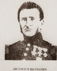
armamentarium of treatments. It was the invention of Antonius Mathijsen, a Dutch
military surgeon in 1851. Essentially, a continuous bandage is wound round and
round the limb then soaked in Plaster of Paris. It is called this because it
was first used extensively in Paris in medicine and in building.
Essentially, it is made from gypsum, which is heated
to produce anhydrous calcium sulphate. When water is added to this it forms
gypsum again and hardens. Hardening takes place very quickly, but it has
to dry out. The larger the cast, the longer it takes. An arm cast will dry out
in 3-6 hours and a leg cast may take up to 6 hours.Yet full drying may not be
complete for 72 hours.
Nikolai Pirogov is credited as the first
surgeon to use them to treat casualties in the Crimean War in the1850s, so of
course, Dr Logan Munro was aware of the method and would use it in his practice
in Wolf Creek!
Diagnosis of fractures
Nowadays we are very dependent on x-rays, but in the
old West there was no such thing. William Roentgen discovered them in 1895.
Only a year later, Dr John Hall-Edwards in Birmingham, England started to use
the so-called X-rays in medical diagnosis.
In the Old West doctors relied on physical examination
to diagnose fractures. Then, and now fractures would be divided into two broad
types:
Closed or simple fractures - when the skin is intact.
Open or compound fractures - when the broken bone
protrudes through the skin.
The type of fracture can vary immensely. We talk about
a 'clean break' meaning a simple transverse fracture. But it can also be
oblique, as can happen with a torsion or twisting injury. Or it can be comminuted, meaning
fragmented. It can be complicated by causing blood vessel or nerve damage. And
it can be a 'greenstick' (as happens in youngsters, when a bone gets bent and
only partially fractures).
The principles of treatment of fractures was
relatively simple then. If moving the part of the body distal to (furthest away
from the site of the pain) caused extreme pain or if grating could be
felt, then a fracture would be diagnosed.
The bone had to be set. That is, the limb had to be
stretched in order to make sure it was the same length as the other. This would
give the best chance to get the two broken ends in position against one another
to allow healing to take place.
Without the use of x-rays it is very likely that all
manner of injuries would end up being diagnosed as a fracture. The treatment
would involve immobilizing the part and hopefully, the injured part would just
'knit together' and be whole again after a few weeks. Remember, that nature
actually does the healing, not the doctor, surgeon or nurse. All that they do
is create the best circumstances for nature to do its job.
And of course if the 'broken bone' healed up all
right, it would bring nothing but kudos to the doctor, whether it actually had
been a fracture or not.
Bone healing
What actually happens when the bone ends are back in
opposition is that a large haematoma, or blood clot forms around the ends. This
is rather like jelly. After a few days blood vessels grow into it and cells
called phagocytes start digesting any debris and tissue that won't heal. Then
other cells called fibroblasts start to lay down collagen, that forms a
framework around the bone ends. This secures the one ends and new bone is
laid down. As a rule of thumb, most bones will have knitted in about 6 weeks.
Some fractures worth knowing about
There are lots of different fractures, many of which
are named after the doctors or surgeons who first described them. Some of the
ones which our western doctors would have known about are as follows:
Colles fracture
A fracture of the distal radius one inch (2.5 cm)
above the wrist. It was described by Abraham Colles an Irish professor of
anatomy in 1814. It is sustained by falling on the outstretched hand as when
you try to break your fall. The problem is that the broken bone gets displaced
and causes a dinner-form abnormality if it is not replaced in posit and
immobilized. It takes 4- 6 weeks to heal.
Smith's fracture
By contrast, a Smith's fracture, also known as a Groyland-Smith fracture is a 'reverse' Colles fracture. It was described by the Irish orthopaedic surgeon Robert Smith in his 1847 book, Treatise on Fractures in the Vicinity of Joints, and on certain forms of Accidents and Congenital Dislocations.
It is a fracture of the distal (bottommost) part of the radius, caused by a blow to the back of the forearm, or a fall on the flex wrist.
Thus, a Colles fracture is an extension injury and the Smith's fracture is a flexion injury.
Bennett's fracture
This is one that could occur in saloon brawls, or
whenever a really hard surface was punched. If you swing at someone and they
duck, causing you to punch the wall, you may end up with a Bennett's fracture.
It can also occur if someone punches someone else's skull! It is a fracture of the first metacarpal, the big bone at the base of the thumb. It is also common in people who have never learned how to punch correctly - which is most people! It was described by Edward Hallaran Bennett, professor of surgery at Trinity College Dublin in Ireland in 1882. It needs immobilization of for 4-6 weeks.
Bennett's fracture
It can also occur if someone punches someone else's skull! It is a fracture of the first metacarpal, the big bone at the base of the thumb. It is also common in people who have never learned how to punch correctly - which is most people! It was described by Edward Hallaran Bennett, professor of surgery at Trinity College Dublin in Ireland in 1882. It needs immobilization of for 4-6 weeks.
Scaphoid fracture
This is not named after anyone, but is the name of one
of the bones of the wrist. It also occurs when you try to break your fall. It
causes pain in the 'anatomical snuffbox.' This is an area at the base of
the thumb. It is called this because in days of yore when folk took snuff, they
placed a pinch in the little depression on the back of the wrist formed when you
elevate the thumb perpendicular to the hand.
The problem with this fracture is
that the scaphoid has a variable blood supply and in many people the blood
supply comes distally. That is, blood vessels do a U-turn to supply the bone
from its far end. If they do not also have a blood supply going directly into
the base of the bone (that is, from the top and bottom) then non-union of the
bone can occur and the piece without the blood supply can die.
The anatomical snuffbox outlined
Monteggia fracture
This is a fracture of the proximal third of the ulna
(the larger bone in the forearm) with dislocation of the head of the radius
(the smaller bone). It was named after Giovanni Monteggi, (1762-1815) an
Italian professor of anatomy and surgery. It occurs with a fall and a twist.
It is a difficult one to treat because the breaks and the two bones are hard to get in the right positions. Nowadays it may necessitate operation.
Monteggia fracture, by Jane Agnes
It is a difficult one to treat because the breaks and the two bones are hard to get in the right positions. Nowadays it may necessitate operation.
Clavicle fracture
The collar bone is commonly injured in contact sports
and fights. A direct blow to the upper chest can fracture it, most
usually at the junction between the middle and outer thirds. They heal up very
well generally. We used to use figure of eight bandages and a sling, but really
they generally just heal without any intervention.
Fracture of neck of femur
The femur is the thighbone. The two main parts are the
neck of the femur, where it forms the hip joint and the shaft, the main part of
the thigh. This is the sort of fracture that happens in older people who
may have osteoporosis, or thinning of the bone. This is nowadays treated by
internal reduction and fixation. It is probably not going to occur to your hero
or heroine in the western novel, but could to another character. It classically
causes a great deal of pain after a fall or twist, and the leg will show
external rotation due to the weight of the limb.
Fracture of the shaft of the femur
This can occur with any large trauma, either directly
from a blow or from a fall. Depending upon which part of the shaft is affected
the muscles will move the two parts ion different directions. The first aid
treatment would be to strap the two legs together, so that the good leg acts as
a splint.
Skull fracture
The big thing here is the possibility of a hemorrhage
inside the skull or into the brain. In terms of the novel, the big question
would be whether to trephine or not. That is, whether to make a hole in the
skull to release blood.
This just happens to be one of the questions that Doc
Logan Munro is forced to consider in Wolf Creek 8: Night of the Assassins.
But you will find no spoilers here!
***
If you are interested in reading more about medicine and surgery in the frontier days, then you may find The Doctor's bag useful. It is a collection of my blog posts, published by Sundown Press.
The novel about Dr George Goodfellow, the Tombstone surgeon to the gunfighters


Keith, as always, a fascinating look into medicine. I can't say enough good about your book, THE DOCTOR'S BAG: MEDICINE AND SURGERY OF YESTERYEAR. That is a "must have" for every historical author, and it's just plain ol' good reading! You have a wonderful way of explaining things in a very interesting way that makes it understandable for those of us who aren't in the medical profession.
ReplyDeleteThank you, Cheryl! I am really pleased that it found a home at Sundown Press.
DeleteAnother interesting post - as always! Like Cheryl says, The Doctor's Bag is a must-have research book for any historical author. I refer to it often. Thanks for all of the fun facts!
ReplyDeleteGood if you to say that J.E.S. The WF blog is fun to write for and I try to make it interesting with little detours and snippets.
DeleteOh those broken bones. I used the plaster cast with my female doctor also, and I checked to make sure she would have known about it first. Now, thanks to this great post, I can write more specifically about this type of injury.
ReplyDeleteI always learn so much from your posts. Thank you. Doris
Thanks, Doris. The process of treating injuries like the Colles fracture can be quite challenging. I still have vivid memories of working in Casualty (now called Accident & Emergency in the UK and the ER in the USA) when I was a junior doctor. You had to manipulate the broken bone into the correct position and hold the limb in the right position while the plaster of plaster was applied. It really is amazing stuff!
DeleteYour book is great and one of the best research tools I have on my desk. Since I'm working on a manuscript where a woman falls down stairs and breaks a leg, this blog came at the perfect time. Thanks.
ReplyDeleteThank you, Agnes, I am pleased that it has been useful.
DeleteClearly a case of synchronicity, here!
Keith, I'm sure I said I have to get your book after reading your previous post but life got a bit hectic these last couple of weeks. I do have a situation I wanted to run by a vet, but perhaps I can e-mail you my question. I loved reading this article and am so glad to be able to get a great reference book. Thanks for posting this valuable information.
ReplyDeleteHi Elizabeth, if I can help, please do email me. dr_keith.souter@btopenworld.com
ReplyDeleteA rich resource for any author, Keith. It motivates me to break one of my character's bones so I can turn to your expertise for a solution.
ReplyDeleteThanks for dropping by, Tom. But please don’t break anybody’s bones on my account!
DeleteKeith,
ReplyDeleteI'm a veteran of several broken bones, which makes this article particularly interesting to me. While I often refer to my copy of "The Doctor's Bag" for medical guidance in my stories, I also enjoy rereading the information when you post your blog articles.
Thanks, Kaye. Practising medicine by clinical impression alone must have been so challenging, back in the days before x-rays. It would be interesting to know whether more or less fractures would have been diagnosed.
Delete